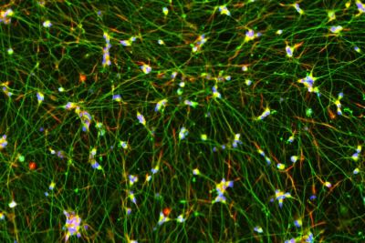
Quick-Neuron™ Excitatory - SeV Kit
| Short Description | This kit differentiates human pluripotent stem cells into excitatory neurons in 10 days using Sendai virus. |
|---|---|
| Product Components | Small: QN-SeV-P, Component N, Component G1, Component G2, Component P, and Coating Material A, Large: QN-SeV-P (undiluted), Component N, Component G1, Component G2, and Component P |
| Storage Conditions | SeV should be stored at -80°C. All other components can be stored at -20°C or -80°C. |
| Product Use | This product is for research use only. It is not approved for use in humans or for therapeutic or diagnostic use. |
| Differentiation Method | Transcription factors delivered by Sendai virus vector |
| Cell Type | Excitatory Neurons |
| Shipping Conditions | Dry ice |
| Shelf Life | 1 year |
Application Notes
Long-term Functional Characterization of Human iPSC-derived Excitatory Neurons with High-Density Multielectode Arrays
Modeling Alzheimer’s disease with ELISA and off-the-shelf patient iPSC-derived Neurons
Human iPSC-derived excitatory neurons and astrocytes differentiated by Quick-Tissue™ technology for functional drug screening on MEAs
Visualizing Neurite Outgrowth by Lentiviral Transduction of Fluorescent Proteins into Human iPSC-derived Excitatory Neurons
Genetic and Functional Profiling of hiPSC-derived Excitatory Neurons Differentiated by Quick-Neuron™ Technology
Posters
Late-stage neuronal development and synaptic maturation of iPSC-derived excitatory neurons (Society for Neuroscience 2023)
In vitro Rett syndrome model based on MeCP2 knockdown in iPSC-derived neurons (Society for Neuroscience 2023)
In vitro Model of Alzheimer’s Disease based on Neurons and Astrocytes Generated from Patient iPSCs using Transcription Factor-Based Rapid Differentiation Technology (Society for Neuroscience 2023)
Epilepsy in a dish (EIDA): a suite of assays to discover therapeutics of epilepsy using isogenic hiPSC-neuron/astrocyte/microglia 2D and 3D tri-cultures and kinetic imaging cytometry (Society for Neuroscience 2023, Vala Sciences)
Establishment of High-Throughput Toxicity Assay System with iPSC-derived GABAergic Neurons Generated by a Rapid Differentiation Method (Safety Pharmacology Society 2023)
Development of Electrophysiological Toxicity Assay System with iPSC-derived GABAergic Neurons Generated by a Rapid Differentiation Method (Safety Pharmacology Society 2023)
Functional and pharmacological evaluation of iPSC-derived astrocytes generated by a rapid differentiation method (Society for Neuroscience 2022)
Functional evaluation of iPSC-derived astrocytes generated by a rapid differentiation method (Neuro2022 in Japan)
Using Human iPSC-derived Neurons and Astrocytes Produced by Transcription Factor-Based Differentiation Protocols to Generate Models to study Alzheimer’s Disease in vitro (Society for Neuroscience 2022)
Generation of Alzheimer’s Disease Model Using Human iPSC-derived Neurons and Astrocytes Produced by a Transcription Factor-Based Differentiation Protocol (Neuro4D 2022)
Separation of drug effects in human iPS cell-derived neurons using MEA system (Society for Neuroscience 2019)
Evaluation of Ready-to-Use Quick-Neuron™ Plate - MEA 48 (Society for Neuroscience 2021)
MEA assay with human iPSC-derived neurons generated by a rapid differentiation method (Safety Pharmacology Society 2020)
Application of Rapid iPSC Differentiation to Neurotoxicity Assays (Safety Pharmacology Society 2020)
User Guides
Quick-Neuron™ Excitatory - SeV Kit (Small)
What kind of transcription factors are used for differentiation induction?
It is a proprietary formulated RNA and cannot be disclosed.
Why do I see many floating cells and/or cellular debris on day 1?
hPSCs were likely damaged while they were harvested. hPSCs may have been mechanically harvested by scraping or were pipetted too much or too vigorously. Lift Solution D1-treated hPSCs only by pipetting them up and down up to 15 times. The mixing of hPSCs with SeV may have also been done too strongly. Gently rock the plate several times to mix hPSCs with SeV.
Why does a reagent in my differentiation kit have less than the required volume?
For reagents supplied in small volumes, be sure to briefly centrifuge before use. If pipettors have been calibrated recently and the volume is less than it is supposed to be even after the tube is spun down, please contact us at [email protected].
What should I do if I find on day 1 that my culture was seeded only in the center but not in the periphery of the well?
Unless cells are accumulated in the center of the well, the instructions can be followed since this is an issue of coating. We have observed that 4-well plates available from Nunc sometimes do not allow cells to settle down in the periphery of the well when our coating conditions are applied. Next time, please use a 24-well plate and try the following: 1) incubate the plate with Coating Material A at 4°C overnight instead of 37°C for 2 hours, 2) do not aspirate the supernatant completely or leave the supernatant just enough to cover the well bottom surface after coating with Coating Material A is completed and 3) do not touch the bottom of wells with fingers when handling the plate.
I plated cells as instructed, so why does my culture look less confluent than the one shown in the user guide on day 1?
hPSCs were likely damaged while they were harvested. Ideally, hPSCs should be lifted only by pipetting several times using Solution D1 as instructed in the user guide. Even if pipetting 15 times does not lift all of the cells in the culture, do not attempt to do more pipetting but collect only cells that come off. Instead rinse the culture with PBS and start Solution D1 treatment again but with longer incubation (up to 20 minutes). Next time please reduce the concentration of the substrate used to coat the dish/well for the maintenance of the source hPSCs to 50-70%.
Why does my culture have colonies that appear balled up and round on day 1?
It could be that Coating Material A was diluted at room temperature or diluted using PBS warmed to room temperature. Keep both Coating Material A and PBS on ice before dilution.
Can I use an orbital shaker for SeV infection while incubating cell suspension for 10 minutes?
The purpose of flicking the tube containing cells and SeV every 2 minutes during the 10 minute incubation is to prevent cells from settling down and forming aggregates. If the orbital shaker can do so, then it is possible to use it.
Can I use Geltrex or Matrigel as a coating substrate for hPSC cultures?
Our kits are not optimized with these substrates. However, if your hPSC differentiation protocols are already optimized with such a substrate, you may try using one of these with our kit at your own risk.
I was able to harvest more hPSCs than I needed for the kit. Can I replate or freeze the leftover hPSCs to maintain their culture?
No. If you’re following the instructions given in the user guide, the hPSCs will be harvested in a differentiation medium so the leftover hPSCs cannot be maintained.
Why are there many aggregates after harvesting hPSCs?
After harvesting hPSCs, cells should not be left in the collection tube for more than 5 minutes. Leaving cells for a longer period may cause the formation of cellular clumps which interferes with infection. Single cells are best for infection.
Why doesn’t Solution D1 dissociate my hPSC culture without scraping?
The concentration of the substrate used to coat the dish/well was likely too high, so Solution D1 treatment requires longer incubation than 10 minutes (up to 20 minutes). Next time, please reduce the concentration of the substrate to 50-70%.
Why do differentiation kits require hPSCs be maintained under StemFit or StemFlex conditions?
hPSCs should be adapted to passaging as single cells prior to differentiation. Our protocols have only been tested with StemFit/StemFlex, so we can't guarantee results using other culture conditions.
How long do I need to maintain hPSCs prior to beginning to use your kit?
Before beginning differentiation, cultures should be healthy and displaying typical morphologies (i.e., round colonies with tightly packed cells exhibiting a small cytoplasmic area), few spontaneously differentiating cells, and normal growth rates (i.e., doubling once a day). We recommend growing the cells for at least 14 days post-thaw before differentiating and not allowing the cultures to reach more than 80% confluency.
Do I need a license agreement for any of Elixirgen Scientific’s products?
No. You don't need any licence or material transfer agreement (MTA) to use our differentiation kits or iPSC-derived cells. However, please be advised that these products are for research use only.
Is Sendai virus (SeV) dangerous?
SeV is not pathogenic to humans (i.e., humans are not the natural host of the virus) and the infection does not persist in immunocompetent animals. Furthermore, SeV used in our kits does not produce infectious viral particles upon transduction to host hPSCs and is a temperature-sensitive mutant, such that it is active at 33℃ but becomes inactive at 37℃. However, because SeV can be transmitted by aerosol and contact with respiratory secretions and is highly contagious, appropriate care must be taken to prevent potential mucosal exposure to the virus. Our SeV-based kits must be used under Biosafety Level 2 (BL-2) containment with a biological safety cabinet or a laminar flow hood and with appropriate personal protective equipment. In the event that the virus comes into contact with skin or eyes, decontaminate the affected area by flushing with plenty of water and follow the safety manual prepared by your laboratory and approved by your Institutional Biosafety Committee.
Will SeV remain active after differentiation?
No. The SeV used in our kits is a temperature-sensitive mutant that is active at 33℃ but becomes inactive at 37℃, which is the temperature instructed in the user guides post-differentiation.
Does Quick-Tissue™ technology leave a genetic footprint?
Sendai virus (SeV) is an RNA virus, so it does not integrate into the genomic DNA. In principle, a foreign gene introduced intracellularly in the form of RNA is quickly translated and expressed because, unlike DNA, RNA does not need to enter the nucleus for forced expression, thereby providing no chance of mutagenesis. This is discussed in the following review paper: Yamamoto, et al., (2009) "Current prospects for mRNA gene delivery." Eur. J. Pharm Biopharm 71, 484-489.
What kind of RNA is used for differentiation induction?
It is a proprietary formulated RNA and cannot be disclosed.


